

At the Ralev Dental Clinic, broken instruments are removed under a rubber dam with the help of a special endodontic microscope that provides good visibility even of the most deeply broken instruments. A broken instrument in the canal is no reason to panic — the decision depends on whether there is inflammation and where the fragment is.
Why does an instrument break in the canal?
Breakage of the tips of the instruments used to remove the pulp (nerve) from the root canals of teeth is a common complication of root canal treatment. It happens for 2 main reasons:
- fatigue of the material the instruments are made of — this occurs through their repeated use;
- difficult and curved root canals of some teeth — they are narrow, hard and curved in the lower 1/3.
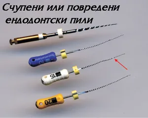
Is a broken instrument dangerous?
Patients are usually very stressed by the fact that there is a broken instrument in their tooth’s canal. This worry is, however, completely unnecessary, because the instruments used in root canals are not in themselves a problem if they stay there. They do not cause allergies, they are not detected by airport metal detectors, they do not cause inflammation, and the material they are made of has no harmful effect on the body.
The problem actually comes from the inaccessible and untreated part of the root canal around and below the instrument.
What can be done about a broken instrument?
In such a situation we have 3 main methods of action: leave it, remove it or bypass it.
First option: the instrument is left in place
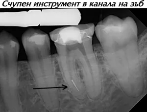
The instrument is left where it is and the root canal treatment is completed without additional manipulation. This option is used in very few situations — mainly when we are sure that there is no infection in the root canal and around the tooth. Such is the case when the tooth is vital and has not caused pain or complaints, but had to be devitalised because of large caries. In that case there is no infection in the root canals, and with precise sterile work under a rubber dam there is no problem if the instrument stays there.
The other common moment when we can afford to leave the instrument is the final stage of filling the canal. At that point the canal has been cleaned and disinfected, and the instrument that breaks is the canal filler we are filling with. It remains included in the tooth’s filling, by analogy with the gutta-percha points that are placed in a standard root canal filling.
This option is applied only and exclusively when we are completely sure of the cleanliness and sterility of the canal, and the canal is filled with a root canal paste that can be easily removed from it.
Such root canals must be monitored with X-rays at 3, 6 and 12 months after treatment. If there are no signs of inflammation and no complaints, the treatment of the tooth is considered complete — there is full function with no clinical or X-ray problems. If, however, signs of inflammation are seen, it is necessary to proceed to the next two options — removing the broken instrument or bypassing it.
Second option: removing the broken instrument
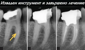
This option is widely known among patients and is due to their fear of the fragment left in the root canal and the thought that it may cause them to lose the tooth. There are many ways to remove a broken instrument, and almost all of them require magnification so that the instrument can be seen during the attempts to remove it. This magnification is provided by microscopes or special magnifying glasses with loupes, according to the individual preferences of the dentist.
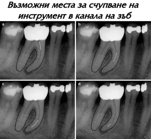
Removal is done using special ultrasonic files, with which the root canal around and above the instrument is widened until it is loosened from the canal wall. Then, using special micro-instruments similar to a cowboy lasso, the instrument is gripped and an attempt is made to remove it. If the attempt is unsuccessful and the instrument does not come out, the canal around it continues to be widened and removal is attempted again.
We usually attempt to remove instruments broken in straight root canals and those located in the upper or middle third of the canal. These are instruments to which we can easily secure visibility and access without removing much of the hard tissue of the root.
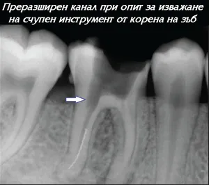
Removing a broken instrument should not be done at any cost, especially for instruments broken in the lower 1/3 of the canal or in curved root canals. With such a “heroic” removal we risk taking away too much of the canal walls, thinning them excessively and thus weakening the tooth, so that the patient later may lose it because of a root fracture during chewing.
If, nevertheless, there is inflammation and the broken instrument prevents us from reaching the untreated part of the canal, but removing it is risky (in thin and curved root canals where we do not have good visibility), another way of dealing with the problem has to be found.
Third option: bypassing
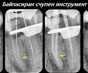
Bypassing means going around the instrument and passing alongside it. In this way we manage to treat the part of the root canal below the instrument and fill it right to the bottom, while the instrument itself stays embedded in the canal wall as part of the tooth and causes no problems. We thus both preserve the walls of the canal without weakening them and eliminate the risk from the unfilled part of the canal.
Bypassing is done with the help of extremely fine instruments with a tip diameter of 0.06 to 0.1 mm, which are used to make a path alongside the broken instrument until it has been fully passed and the original canal is reached. The procedure is done exclusively by hand, relying on the dentist’s sense of touch. Using magnification helps, but is not mandatory.
Another specific feature of bypassing is that it takes quite a lot of time — sometimes several visits may be needed to get past the broken instrument. There are patients in whom, after bypassing, we also manage to remove the instrument from the root canal. In this case we are maximally micro-invasive and have done the most possible for the tooth.
What is done if the three methods do not help?
There are teeth in which these 3 methods do not give a result — we cannot remove or bypass the instrument and there is inflammation present. Then there are other ways of dealing with the problem — they are microsurgical. In them, only the tip of the root where the instrument is broken, or the whole root, is removed and the inflammation around it is cleaned out under visual control.
These methods, although surgical, give a more lasting, faster and easier resolution of the problem in certain teeth, and are called apical osteotomy and hemisection (read more).
Frequently asked questions
Is a broken instrument in the canal dangerous?
Not in itself. The instruments do not cause allergies, they are not detected by airport metal detectors, they do not cause inflammation and their material has no harmful effect on the body. The problem comes from the inaccessible and untreated part of the canal around and below the instrument.
Why do instruments break in root canals?
For 2 main reasons: fatigue of the material through repeated use, and difficult, narrow, hard and curved root canals in the lower 1/3.
Is a broken instrument always removed?
No. There are 3 options: the instrument can be left, removed or bypassed. Removal is not done at any cost, because for instruments in the lower 1/3 or in curved canals it may thin the root walls excessively and weaken the tooth.
What is bypassing a broken instrument?
The instrument is bypassed with extremely fine instruments, the part of the canal below it is treated and filled, and the instrument itself stays embedded in the canal wall. It takes quite a lot of time and sometimes several visits are needed.
How does the clinic work when removing a broken instrument?
Under a rubber dam and with an endodontic microscope, which provides good visibility even of the most deeply broken instruments.